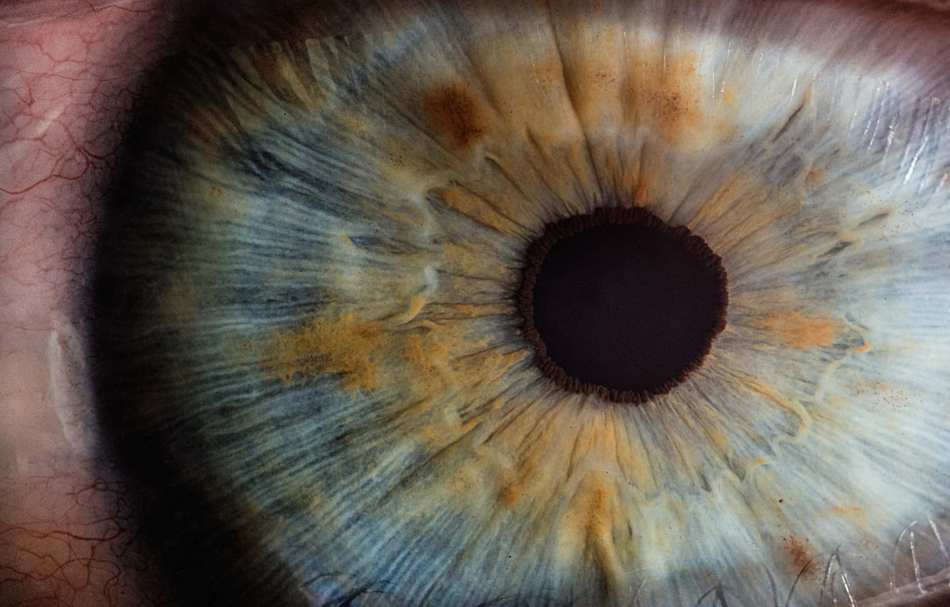
Glaucoma | 青光眼
- Primary glaucoma | 原发性青光眼
- Primary open-angle glaucoma, POAG | 原发性开角青光眼
- Primary angle closure glaucoma, PACG | 原发性闭角青光眼
- Secondary glaucoma | 继发性青光眼
IOP
- 眼压是青光眼发生发展的最重要危险因素
- 降眼压是目前唯一获得证实的可以减缓青光眼疾病进展和保留视功能的治疗方法
- 眼压和眼压测量仍是青光眼诊断与治疗中最基本、最重要的指标
- 较高的眼内压及 MD 导致较高的失明风险
Intraocular pressure 与 blood pressure 的关系
参考文章:Ocular perfusion pressure and glaucoma: clinical trial and epidemiologic findings
眼灌注压表示为动脉血压与眼内压差值
Perfusion pressure | 灌注压
The normal functioning of tissues depends on the maintenance of an adequate perfusion, with sufficient blood flow. A key element is the presence of an ample perfusion pressure to meet tissue needs, a process that requires a balance between arterial and venous blood pressure (BP).
人体组织器官需要足够的血流量才能正常工作,血流量与灌注压直接相关。
Ocular perfusion pressure, OPP | 眼灌注压
参考:The Role of Ocular Perfusion Pressure in Glaucoma
OPP 定义为眼内动脉压与静脉压差值。眼内静脉压接近 IOP,因此可使用 IOP 进行近似,代替静脉压:
$$ OPP = arterial \ BP - IOP $$但动脉压要复杂一些,动脉压由心脏跳动产生,存在收缩压(systolic BP)、舒张压(diastolic BP)和平均血压的概念,因而引入了三个计算公式:
$$ Systolic \ OPP \ (SPP) = Systolic \ BP - IOP $$$$ Diastolic \ OPP \ (DPP) = Diastolic \ BP - IOP $$$$ Mean \ OPP \ (MPP) = \frac{2}{3} \times [diastolic \ BP + \frac{1}{3} \times (systolic \ BP - diastolic \ BP)] - IOP $$Low OPP 是青光眼的明确危险因素
Low DPP is a risk factor for glaucoma.
— Jeffrey Liebmann, MD
已在统计学上建立了 BP 与 IOP 关系
Many studies have reported a positive, statistically significant relationship between blood pressure (BP) and IOP, which is consistent across studies and stronger for systolic than for diastolic BP. Additionally, BP often explains a larger proportion of the variation in IOP than other known variables, although the percent explained is relatively small, about 5%. For systolic BP, estimates from cross-sectional studies suggest that each 10 mmHg higher BP is associated with a 0.23−0.32 mmHg higher IOP. Similar estimates, of 0.21 and 0.22mmHg higher IOP, have been derived from longitudinal data in the Beaver Dam and Barbados studies. Results for diastolic BP are less consistent.
关键点:
- BP 与 IOP 在统计学上呈正相关;
- BP 是已知能解释 IOP 变化的最显著变量;
- 收缩压与 IOP 相关性强于舒张压;
- 收缩压每升高 10 mmHg,眼压上升 0.2 - 0.3 mmHg;
OPP 与 IOP 的联系有待研究
The transient IOP fluctuations (IOP minus IOP baseline) are positively correlated with the ocular perfusion pressure (mean blood pressure minus IOP)
OPP 与 IOP 波动成正相关,这个是显然的。
A low ocular perfusion pressure could be due to a relatively low BP or a relatively high IOP. Neither of these variables, alone, has been implicated conclusively as the sole cause of low perfusion in glaucoma. As discussed in a later section, there is no clear relationship between BP levels and OAG damage.
高 IOP 或低 BP 都可以得到低 OPP,但这个看起来有点类似鸡生蛋的问题。
角膜力学性能测量
Corneal biomechanical properties are important for the diagnosis of corneal diseases, individualized design and prognosis of corneal surgery. Clinical available devices such as Ocular Response Analyzer (ORA) and Corneal Visualization Scheimpflug Technology (Corvis ST) can provide corneal biomechanics related parameters, while corneal elastic modulus cannot be extracted directly from them at present.
角膜生物力学特性对于角膜疾病的诊断、角膜手术的个体化设计和预后具有重要意义。临床可用的设备如眼反应分析仪(ORA)和角膜可视化Scheimpflug技术(Corvis ST)可以提供角膜生物力学相关参数,但目前无法直接从中提取角膜弹性模量。
Ocular pulse | 眼脉搏
The ocular pulse amplitude is defined as the difference between diastolic and systolic intraocular pressure. The ocular pulse is generated by the pulsatile ocular blood flow in the choroid. It is dependent on the dynamics of the cardiovascular system, the rigidity of the ocular vessels on one side and the biomechanical properties of the eye on the other side. 眼脉搏幅度定义为舒张压和收缩压眼压之间的差值。眼脉搏是由脉络膜中脉动的眼部血流产生的。它取决于心血管系统的动力学、一侧眼血管的刚性以及另一侧眼睛的生物力学特性。
Dynamic contour tonometry (Pascal®) does not only measure intraocular pressure almost independent of corneal thickness and curvature but also allows easy and fast measurement of ocular pulse amplitude on the slit lamp. 动态轮廓眼压计 (Pascal®) 不仅可以测量几乎与角膜厚度和曲率无关的眼内压,还可以在裂隙灯上轻松快速地测量眼脉搏幅度。
The ocular pulse amplitude in healthy subjects is between 1.2 and 4 mmHg. If the ocular pulse amplitude is larger than 1.2 mmHg spontaneous pulsations of the central retinal vein are visible on fundoscopy. 健康受试者的眼脉搏幅度在 1.2 至 4 mmHg 之间。如果眼脉搏幅度大于 1.2 mmHg,则在眼底镜检查中可以看到视网膜中央静脉的自发搏动。
In patients with ocular hypertension the ocular pulse amplitude is larger than in normal subjects but this is mainly due to higher IOP levels. 高眼压患者的眼脉搏幅度比正常人大,但这主要是由于眼压水平较高。
— J P E Stürmer3
参考书籍
Becker-Shaffer’s Diagnosis and Therapy of the Glaucomas
Here is Prof. Lin Li’s profile page. ↩︎
Evaluation of corneal elastic modulus based on Corneal Visualization Scheimpflug Technology. ↩︎
Role of ocular pulse amplitude in glaucoma Article in German. ↩︎